
Corpus Christi
2701 Morgan Ave #425, Corpus Christi, TX 78405


An image-guided procedure that uses radiofrequency energy to create a controlled lesion on selected medial branch nerves, considered for facet-mediated axial neck or low back pain after qualifying diagnostic blocks and clinical review.
Two audiences read this page: the patient considering treatment and the professional coordinating the related care or claim.
Patients with predominantly axial neck or low back pain whose clinician is considering RFA after qualifying diagnostic medial branch blocks.
Firms coordinating a spine-injury claim that need the indication, diagnostic-block response, treated levels, procedure details, and follow-up plan documented clearly.
Radiofrequency ablation (RFA) uses radiofrequency energy to create a controlled thermal lesion on selected medial branch nerves. The goal is to interrupt pain-signal transmission from facet joints evaluated through the clinical record and qualifying diagnostic blocks.
A response to diagnostic blocks can support patient selection, but it does not guarantee the outcome of RFA. Benefit, duration, and recovery vary, and nerves can regain function over time. The treating clinician applies current clinical and payer criteria to each case.
RFA may be considered when the clinical picture and qualifying diagnostic blocks support a facet-mediated pain pathway.
The indication, target levels, preparation, and follow-up plan are confirmed before the procedure.
The clinician reviews symptoms, examination findings, imaging, prior care, diagnostic-block responses, and applicable criteria.
The patient is positioned, the treatment area is cleaned, and local anesthetic is used according to the clinical plan.
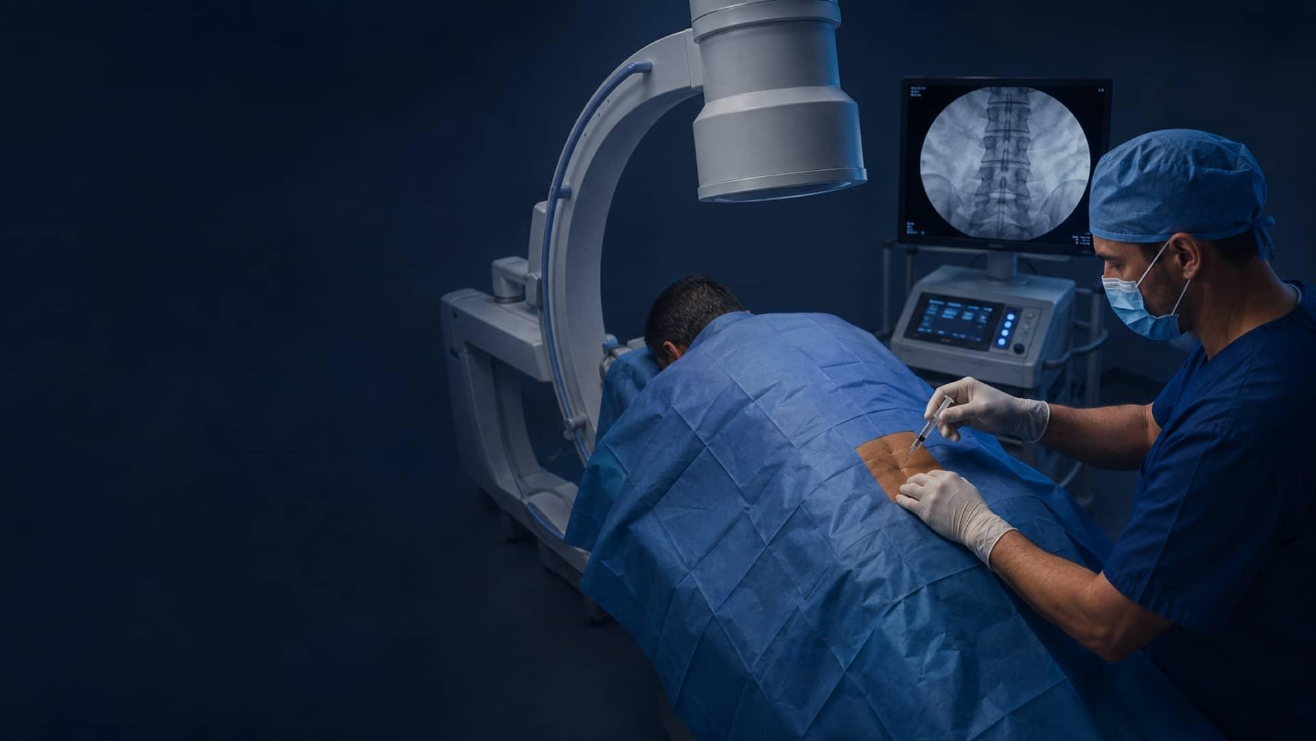
The physician uses fluoroscopic imaging to guide specialized needles toward the selected medial branch targets.
Stimulation may be used when clinically appropriate before controlled radiofrequency energy creates the planned thermal lesion.
The patient is observed before discharge and follows individualized activity, medication, symptom-monitoring, and follow-up instructions.
Preparation and follow-up vary by target region, medical history, medication, facility instructions, and payer criteria.
The team confirms check-in, medication, fasting, and transportation instructions before the appointment.
Fluoroscopy is used to guide placement at the selected medial branch targets.
The degree and duration of benefit vary, and improvement is not guaranteed.
Follow-up timing, activity progression, and further treatment are individualized.
The clinician reviews the treatment goal, alternatives, limitations, and patient-specific risks before treatment.
GTOA coordinates referral intake, scheduling, available records, and status communication for patients and authorized legal teams.
History, mechanism of injury, examination findings, diagnosis, and the proposed treatment plan.
Available imaging is considered alongside symptoms, examination findings, prior care, and alternative pain sources.
When clinically appropriate, documentation may discuss how the reported incident, findings, and pre-existing conditions relate.
Indication, treated levels, guidance, testing when used, lesion parameters, tolerance, and follow-up are documented as appropriate.
The clinician may document rehabilitation, follow-up, repeat treatment, or a different pathway when supported by the record.
Scheduling and records timing depend on location, clinical need, authorization, record availability, and clinician review. The team confirms the next step for each request.
Coverage and payment arrangements vary. Contact the team before scheduling so eligibility, authorization, and current options can be confirmed.
Contact GTOA to verify current plan participation, benefits, and any authorization requirements before the procedure.
Lien-based arrangements may be available for qualifying personal injury cases. The team confirms eligibility and terms for each referral.
Patients paying directly can contact the office for current pricing and available payment arrangements.
The same six reasons matter to a patient choosing a clinic and to a firm choosing where to send a client.
Symptoms, examination findings, imaging, prior care, diagnostic responses, and alternative pain sources are reviewed before RFA is recommended.
Radiofrequency ablation is performed with fluoroscopic guidance at the selected medial branch targets.
Diagnostic-block responses and current clinical or payer criteria are considered before treatment.
Documentation can identify the indication, treated levels, approach, guidance, testing, lesion parameters, and follow-up plan.
Patients and authorized legal teams can contact GTOA about scheduling, status questions, and available records.
Beaumont, Houston, San Antonio, Dallas, Arlington, and Corpus Christi provide access to GTOA care across Texas.
Patients can request care through Contact Us, and legal teams can begin a referral through the For Attorneys page.
The physician responsible for the medical review of this page.
Six clinics across Texas. Choose the one closest to you for hours, phone, and directions.
2701 Morgan Ave #425, Corpus Christi, TX 78405

907(A) Medical Center Drive, Arlington, TX 76012

8330 Lyndon B Johnson Fwy, Suite B400, Dallas, TX 75243

7220 Louis Pasteur Drive, Suite 130, San Antonio, TX 78229

8928 Kirby Drive, Houston, TX 77054

324 North 23rd St, Beaumont, TX 77707
Services directly connected to diagnostic selection for RFA or to evaluation when another spine pain source is identified.
The clinician considers symptoms, examination findings, imaging context, conservative care, diagnostic-block responses, procedural accuracy, and applicable clinical or payer criteria.