Loading article


A sore shoulder after a fall, a swollen ankle after a twist, or a sudden loss of strength can point to several injuries. The next question is often whether a tendon or ligament is irritated, partially torn, completely disrupted, or moving abnormally. Musculoskeletal ultrasound can help answer it while the clinician examines the painful area in real time.
This test uses sound waves rather than ionizing radiation. A transducer sends sound into the body and converts returning echoes into moving images. For many superficial tendons and ligaments, ultrasound may show fiber continuity, fluid, tendon or ligament disruption, retraction, and movement during dynamic testing. Its value depends on the structure, the clinical question, and the examiner’s experience.
Musculoskeletal ultrasound works best when it is tied to a defined clinical target. A clinician first considers how the injury occurred, where tenderness is located, which movements reproduce symptoms, and whether strength or stability has changed. Imaging then adds information the examination cannot provide with enough confidence.
The scan may help distinguish among several findings that can feel similar to a patient:
Every comment is reviewed before publication. Do not include medical details, personal identifiers, or information about an active claim. This form is not monitored for medical questions or emergencies.
Three more from the same thread.
Showing the first 3 of 3 articles
These findings can help guide protection, rehabilitation, referral, or additional imaging, but they should be interpreted alongside the injury history, examination, and functional loss.
During an orthopedic evaluation, the clinician may test motion, strength, tenderness, stability, and the ability to perform a specific task. That information gives the examiner a target. Without it, a detailed scan may identify changes unrelated to the patient’s symptoms or miss a problem outside the scanned region.
Imaging findings and pain do not always match. Tendon thickening may be chronic, and a small defect may be important in one location but incidental in another. The report must be interpreted with the injury history and examination.
Ultrasound for tendon injuries can evaluate common problems involving the rotator cuff, biceps, Achilles tendon, patellar tendon, quadriceps tendon, and tendons around the elbow, wrist, hip, and ankle. Interpretation depends on probe angle and technique because anisotropy can make a normal tendon appear abnormally dark. Injury can alter that pattern, create a gap, produce surrounding fluid, or change the way the tendon glides.
The examiner can scan along and across the tendon, measure a defect, and compare sides when useful. With ultrasound for tendon injuries, movement adds information a still image cannot provide. Watching the tendon during joint motion or muscle contraction may reveal snapping, subluxation, impingement, or separation of torn ends.
Acute swelling and healing can change the appearance over time, so follow-up imaging may be useful when the result could affect care.
Ultrasound for ligament injuries is best suited to structures that can be reached and visualized from the skin surface. The examiner looks for fiber disruption, thickening, surrounding fluid, abnormal laxity, or a small avulsion near the ligament attachment. When clinically appropriate, controlled stress may help assess abnormal laxity or widening across a ligament-supported joint.
This approach is useful for the lateral ligament complex of the ankle, the ulnar collateral ligament at the elbow, selected wrist and hand ligaments, and the collateral ligaments of the knee. Still, ultrasound for ligament injuries is not equally effective for every structure. Ligaments deep inside a joint can require MRI or another test for a complete assessment. An uncertain scan should not overrule strong clinical evidence.
The injured area is positioned for safe access and movement. Gel is placed on the skin, and the transducer passes over the suspected structure from more than one direction. The patient sometimes changes position, contracts a muscle, or moves the joint. Gentle probe pressure can identify focal tenderness or shifting fluid.
Musculoskeletal ultrasound requires no radiation or enclosed scanner. Brief discomfort is possible when the probe passes over a tender area, but the examination itself is noninvasive. Examination time varies with the number and complexity of structures assessed.
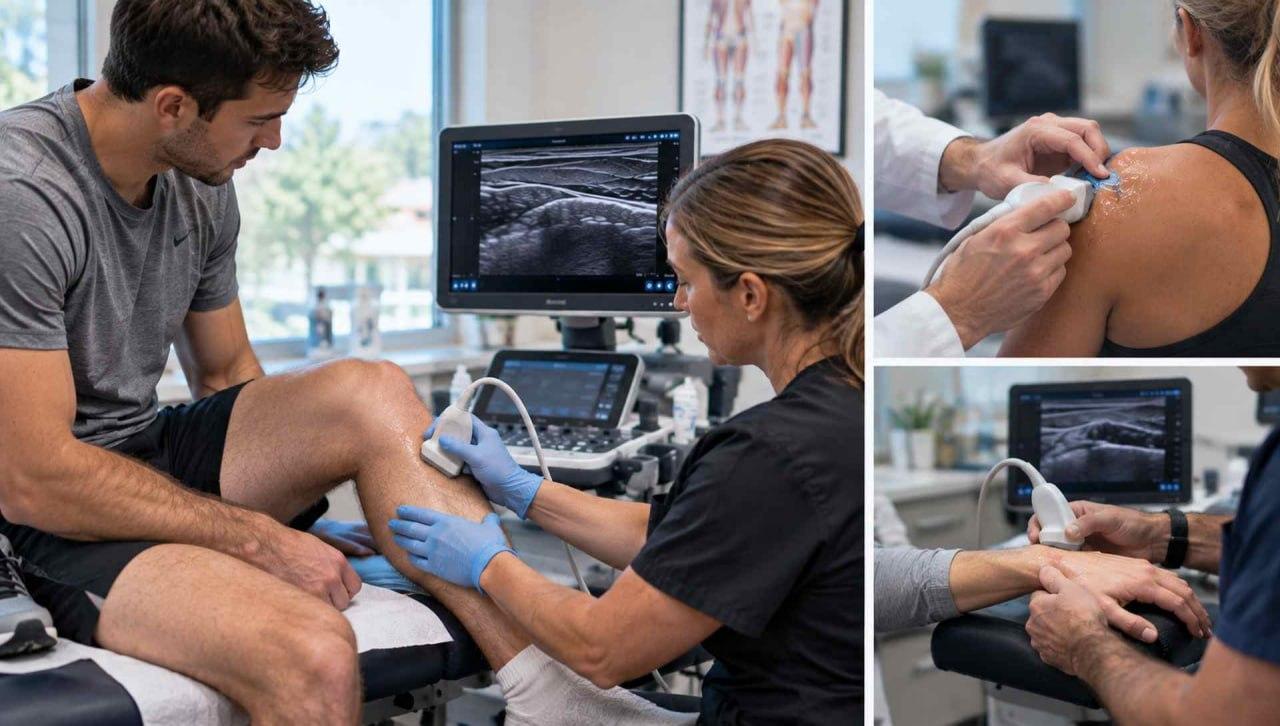
MRI produces detailed images with the patient usually remaining still. Ultrasound offers a different kind of information. The clinician can watch a tendon slide, test a ligament under controlled stress, or reproduce a snapping sensation while viewing the structure involved. The painful movement and the image can be correlated at the same moment.
Guarding, pain, stiffness, and technique can affect what the examiner sees. Dynamic testing should remain controlled and should not be forced when a major acute injury is suspected.
A useful report identifies the structure and side, the location and dimensions of an abnormality, dynamic findings, and nearby changes. For a visible tear, it describes whether the defect appears partial or complete and whether there is retraction. Saved images permit later review.
The injury mechanism, symptom onset, examination findings, and effect on function explain why imaging was obtained. Objective imaging can strengthen the medical record, but it cannot independently establish when an injury occurred or explain every reported limitation.
When patients compare musculoskeletal ultrasound vs MRI, the useful issue is which test can resolve the suspected problem. Ultrasound is useful for selected superficial and dynamic problems, while MRI may be better for deeper or more complex injuries.
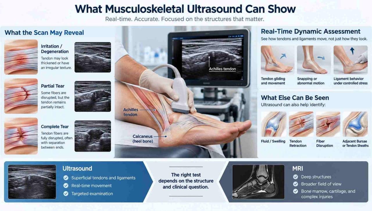
The ultrasound examination can compare sides, focus on the area of tenderness, and change the viewing angle during the examination. It can guide selected injections or aspirations because the needle and nearby tissues are observed during the procedure. These advantages are strongest for superficial targets in trained hands.
A particular strength of ultrasound for tendon injuries is the combination of detailed surface imaging and movement. It can show whether tendon fibers remain continuous and whether a suspected tear changes during contraction. The examination can also assess adjacent bursae, tendon sheaths, fluid collections, and some superficial nerves when clinically relevant.
MRI provides a larger field of view and depicts deep structures that sound waves do not reach well. Bone blocks ultrasound, and tissue depth can reduce image quality. MRI often adds information about internal joint structures, occult bone injury, marrow, cartilage, or complex injury patterns.
The choice of musculoskeletal ultrasound vs MRI is sometimes sequential. An inconclusive ultrasound can still narrow the possibilities and show why another test is needed. The reverse can also occur. A targeted dynamic scan can explain a symptom that was not reproduced during a static MRI. Readers who want more context can review how MRI findings are interpreted after an injury.
Ultrasound accuracy depends on technique, probe angle, operator experience, body habitus, tissue depth, and the structure being examined. Some injuries cannot be fully assessed with ultrasound alone, and additional imaging may be needed when the clinical concern remains.
Imaging becomes useful when it changes a decision. An injury with preserved strength and stability is often managed with protection, activity modification, rehabilitation, and follow up. A complete tear, substantial retraction, instability, or significant loss of function may warrant prompt specialist evaluation. The image remains one part of that decision.
Seek urgent medical attention after an injury if there is an obvious deformity, an open wound, rapidly increasing swelling, severe weakness, new numbness, or a hand or foot that becomes pale or cold. These findings may indicate a problem that should not wait for a routine imaging appointment.
Patients can ask which structure explains their symptoms, whether the finding appears acute or chronic, how fully the injury was seen, and whether another test would change treatment. They should also ask which activities are safe before the next visit.
Final Thoughts
Useful imaging starts with a careful examination and a defined clinical target. When ultrasound can assess the suspected tendon or ligament, it provides detailed information during movement. When depth, bone, or the extent of injury leaves uncertainty, additional imaging can close the gap. The result matters most when it leads to appropriate protection, rehabilitation, referral, or follow up for the individual patient.
Still Having Pain or Loss of Function After a Tendon or Ligament Injury?
Greater Texas Orthopedic Associates can evaluate persistent pain, weakness, swelling, instability, or loss of function and help determine whether musculoskeletal ultrasound or other imaging may be appropriate.
Musculoskeletal ultrasound can identify tendon or ligament thickening, fluid, partial tears, complete tears, retraction, and abnormal movement in structures that are accessible to the probe.
No. Musculoskeletal ultrasound uses sound waves to create real-time images and does not use ionizing radiation. The examination is generally noninvasive, although pressure from the probe may feel uncomfortable over a tender area.
Neither test is always better. Ultrasound is especially useful for superficial structures and dynamic movement assessment, while MRI may provide more information about deep tissues, bone marrow, cartilage, and complex joint injuries.
Your clinician reviews the ultrasound images together with your symptoms and physical examination before recommending treatment, rehabilitation, follow-up, or additional testing if needed.
Medical disclaimer: This article provides general educational information and does not replace an individual medical evaluation, diagnosis, or treatment plan.
Comments are reviewed before they appear. For appointments, records, or medical questions, use the contact page.
No comments have been published on this article yet.