Loading article
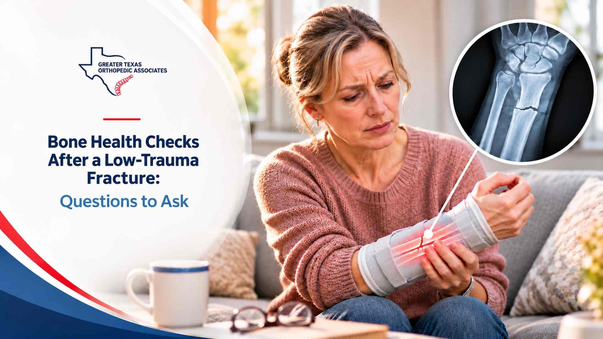
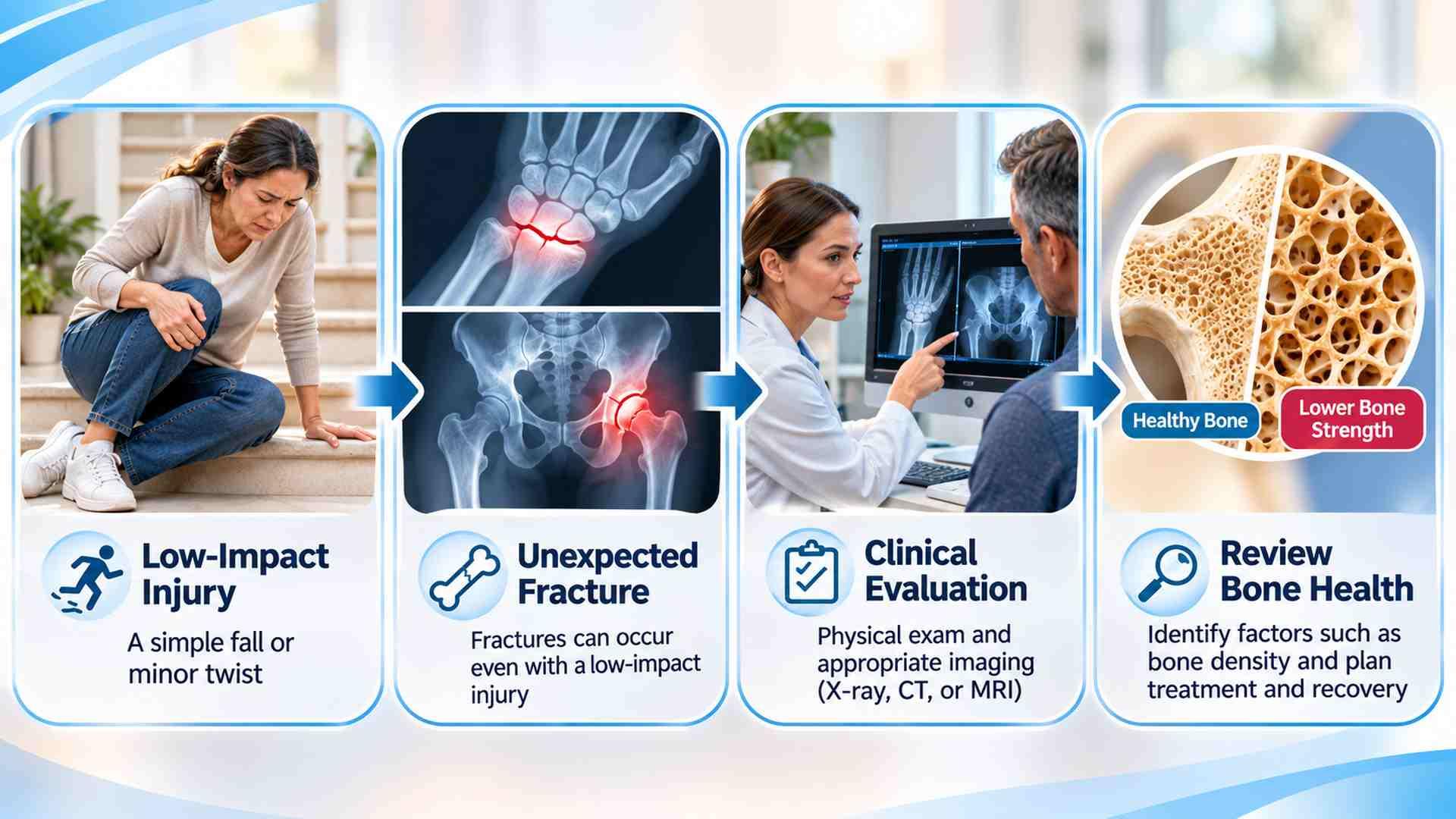
A wrist breaks after a trip on a level sidewalk. A vertebra fractures while lifting something light. After immediate treatment, ask why the bone broke under that amount of force. A bone health assessment after fracture can identify reduced bone strength, contributing conditions, and steps to reduce the risk of another break.
This discussion is especially relevant after age 50, although an unexpected break in a younger adult also deserves consideration. Use these questions at your next visit. This article provides general education and does not replace individual medical evaluation.
A fragility fracture generally follows force that would not ordinarily break healthy bone, often a fall from standing height or less. The hip, spine, wrist, pelvis, and upper arm are important sites. However, the location and circumstances matter: a broken finger after a direct blow does not carry the same implications as a hip fracture after a simple trip.
“Does the force involved explain this break, or should we investigate bone weakness?” Describe the fall height, how you landed, and whether pain preceded the incident. A low trauma fracture evaluation considers these details alongside age, fracture pattern, and medical history; circumstances alone do not establish the cause.
Mention dizziness, blackouts, previous falls, and difficulty rising from a chair; these may need assessment beyond the injured bone. Bring reports of earlier fractures, height loss, family hip fractures, or osteoporosis, including results described as mildly abnormal.
“Would a bone density test after fracture change my care?” is a useful starting question. Central dual-energy X-ray absorptiometry, called DXA or DEXA, usually measures mineral density at the hip and lower spine. It uses a small amount of radiation and helps assess future fracture risk. It does not determine whether the current break has healed.
Every comment is reviewed before publication. Do not include medical details, personal identifiers, or information about an active claim. This form is not monitored for medical questions or emergencies.
Three more from the same thread.
Showing the first 3 of 3 articles
A bone health assessment after fracture differs from routine screening in someone without symptoms or an injury. The fracture itself may justify investigation before the usual screening age. This applies to men as well as women; being younger than the routine screening threshold does not settle whether testing is appropriate.
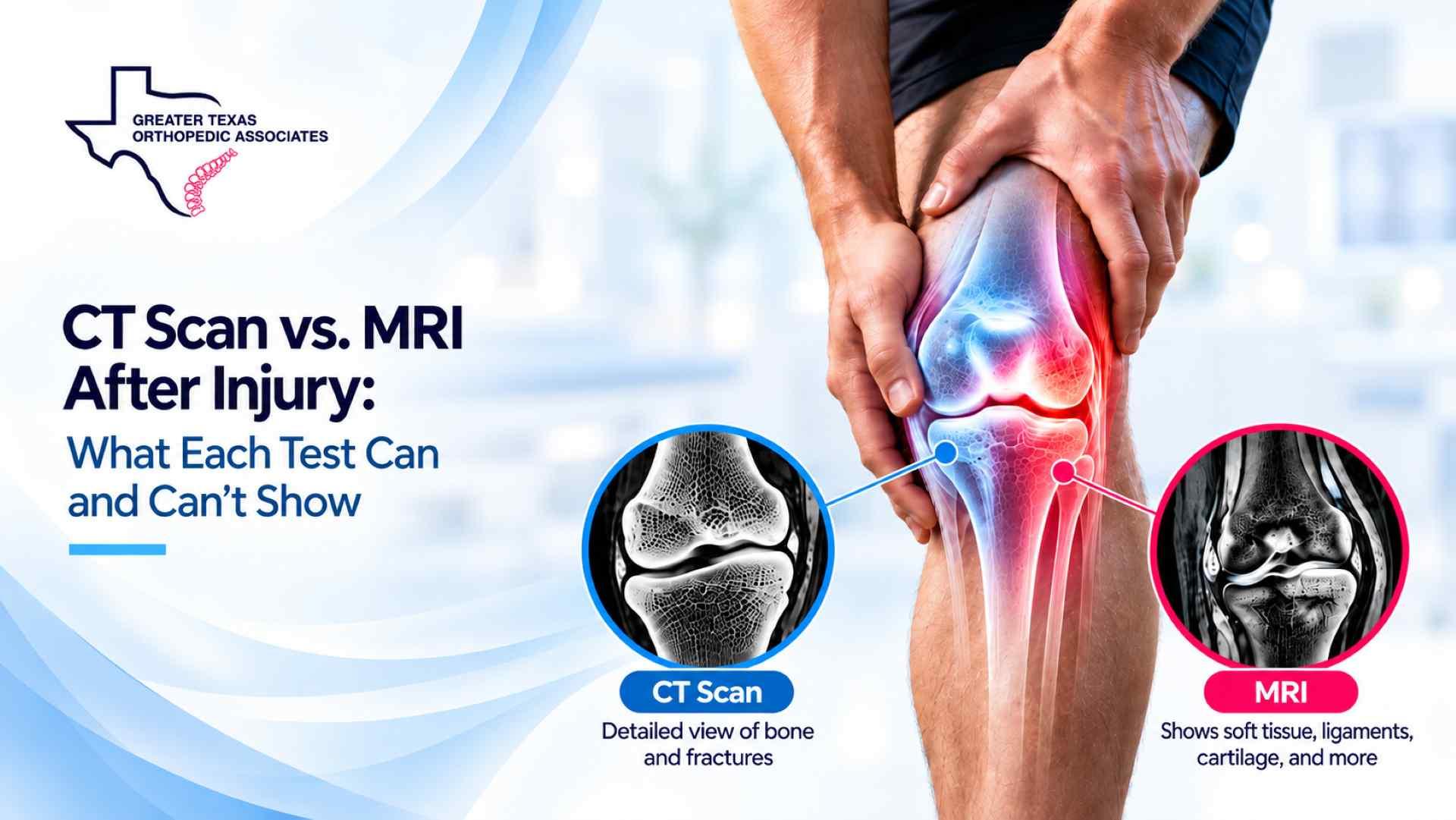
Injury imaging answers a different question. GTOA’s explanation of CT and MRI after injury describes their roles in assessing bone and soft tissue damage. Routine injury scans do not automatically replace formal density assessment, although findings may warrant further bone evaluation.
For postmenopausal women and men aged 50 or older, clinicians generally interpret T-scores. A value at or below minus 2.5 at a valid hip or lumbar spine site meets the bone density criterion for osteoporosis. Before menopause and in men younger than 50, Z-scores are usually preferred; these compare density with an age-matched population. A low result in a younger adult needs interpretation in the context of fractures and possible underlying disease.
“Were the measured areas reliable?” Arthritis, surgery, or implants can affect interpretation. The report should identify usable sites and limitations. Mention pain that makes lying still difficult so the imaging team can plan support.
“Could a condition or medicine have weakened my bones?” Possible contributors include prolonged prednisone use, thyroid or parathyroid disorders, kidney disease, poor nutrient absorption, and low sex hormones. Mention early menopause, weight changes, restrictive eating, and digestive problems.
A low trauma fracture evaluation may include blood tests for calcium, kidney function, vitamin D, and other findings guided by your history. Additional hormone tests or investigations for absorption problems are appropriate for selected patients. Blood work looks for contributors and helps guide safe treatment; a normal calcium level does not prove that bone strength is normal.
If you are being assessed through GTOA’s injury care departments, mention conditions that could affect bone strength. Ask who will investigate them or recommend a specialist, and who will review results from different practices together.
Bring prescriptions, supplements, doses, and duration of use. Some medicines affect bone; others increase falls through dizziness or sedation. Do not stop prescribed steroids or essential treatment yourself. Discuss alternatives with the prescriber. Note previous osteoporosis treatment, missed doses, and side effects.
“How would my risk estimate change the plan?” FRAX combines personal risk factors, sometimes with hip density, to estimate the chance of a hip or other major osteoporosis-related fracture over ten years. The result helps inform treatment alongside your fracture history.
The calculator accepts ages 40 to 90 and uses country-specific models. In U.S. osteoporosis care, its treatment thresholds generally guide postmenopausal women and men aged 50 or older. Recent fractures or repeated falls can require a different clinical judgment than the number alone suggests.
A reassuring density result does not erase the significance of a fragility fracture. In postmenopausal women and men aged 50 or older, a hip or vertebral fragility fracture can warrant osteoporosis treatment regardless of bone density. Decisions after other fracture types depend on the overall assessment. Ask what finding supports the recommendation rather than assuming everyone needs the same medicine.
Some vertebral fractures cause little pain and go unrecognized. Ask whether height loss or stooping could reflect an older spine fracture. Spine imaging or vertebral fracture assessment with DXA may be appropriate. Identifying an old break can change treatment even when the current injury involves your wrist.
“Should prevention treatment begin before my fracture has healed?” Raise this during your follow-up. When medication is indicated, discuss expected benefit, kidney function, side effects, and how treatment will be monitored or changed. Waiting for a scan should not automatically delay necessary prevention; the treating clinician determines what can start safely.
Calcium, vitamin D, and adequate protein support bone health, but supplements alone may be insufficient when fracture risk is high. Review your food intake before deciding whether supplements are needed. Avoid choosing large doses without advice, particularly with kidney disease or a history of stones. Prevention also includes smoking cessation and reviewing alcohol intake.
Exercise must respect the current fracture. Ask when weight bearing, resistance work, and balance training can progress. A therapist can help with transfers, stairs, and walking aids. Review loose rugs, lighting, footwear, vision, and medicines that cause unsteadiness to address fall risk.
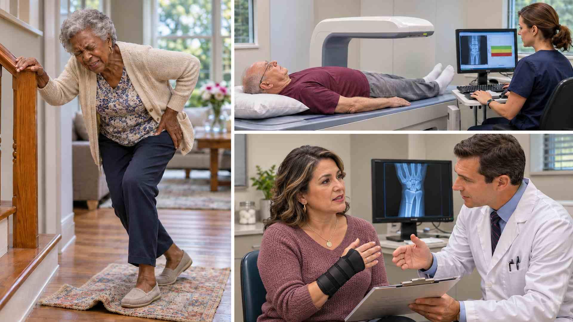
The fracture clinician, primary care clinician, and osteoporosis specialist may have different roles. A fracture liaison service, where available, can coordinate prevention. Confirm who will arrange tests, review results, and explain treatment recommendations.
Bring a short written record so the next clinician can see what has already been assessed. Leave space beside your questions to record the agreed plan. Useful details to have ready include:
For follow-up through Greater Texas Orthopedic Associates, explain which records are available and which evaluations remain outstanding. Confirm where results will be sent and who will explain them. If testing is recommended, follow up if scheduling or the results discussion does not occur.
Agree when the prevention plan will be reviewed. Repeat density testing needs a purpose and an interval suited to your treatment and risk; using the same facility helps comparison. A new fracture during treatment warrants reassessment before the next scheduled scan.
Routine testing must not delay emergency assessment. Seek emergency care for a cold or pale injured limb or an open wound over a suspected fracture, even if bone is not visible. Back pain with new bladder or bowel dysfunction, numbness around the groin, or progressive leg weakness also needs emergency assessment. New focal back pain or worsening fracture pain and swelling warrants prompt clinical advice.
Before leaving, confirm whether testing is needed, what can begin now, and who will explain results. If testing or treatment is deferred, ask why and when that decision will be reviewed. Write down the next step.
Not necessarily. The fracture location, force involved, medical history, and testing help your clinician assess bone strength.
Your clinician may recommend DXA based on your fracture and risk factors, even before routine screening would begin.
They can identify contributing conditions or deficiencies. Normal blood calcium alone does not establish that your bones are strong.
Confirm which clinician will order tests, explain results, review medicines, and coordinate fracture prevention while your injured bone continues healing.
Comments are reviewed before they appear. For appointments, records, or medical questions, use the contact page.
No comments have been published on this article yet.