Loading article
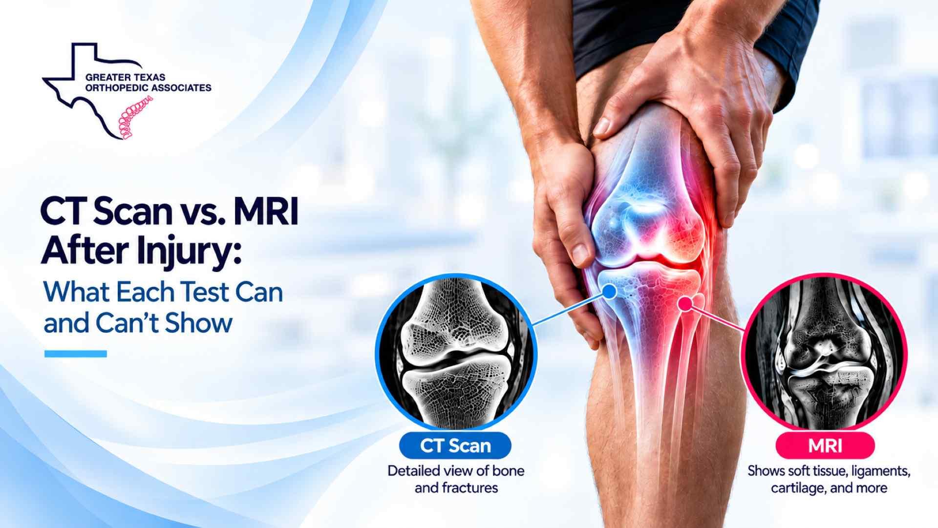
CT and MRI can both help investigate an injury, but they answer different questions. CT is particularly useful for showing the shape and position of broken bone. MRI often provides more information about ligaments, tendons, cartilage, discs, and changes within bone marrow. Choosing a CT scan vs MRI after injury depends on the suspected problem and how the result would change your care.
For example, an ankle fracture that extends into a joint may need a different imaging approach from an ankle that keeps giving way despite a normal X-ray. The examination, symptoms, and existing images help your clinician decide whether another scan is needed and which study is likely to be useful.
| Question | CT scan | MRI |
|---|---|---|
| What does it use? | X-rays and computer processing | Magnetic fields and radiofrequency signals |
| Where can it be especially helpful? | Fracture patterns, bone fragments, and joint-surface detail | Ligaments, tendons, cartilage, discs, bone marrow, and selected hidden fractures |
| What are its limitations? | Routine CT may not explain some ligament, tendon, or meniscal injuries | Findings may be incidental or pre-existing; MRI is not automatically the preferred test for a complex fracture |
Every comment is reviewed before publication. Do not include medical details, personal identifiers, or information about an active claim. This form is not monitored for medical questions or emergencies.
Three more from the same thread.
Showing the first 3 of 3 articles
| Does it use ionizing radiation? | Yes | No |
| What is the experience like? | Generally quicker, though the examination and preparation vary | Usually involves lying still for longer in a loud scanner; implant screening is necessary |
These are general strengths, not a rule that CT only shows bone or MRI only shows soft tissue. The body part, scan protocol, and clinical question matter. In selected cases, both tests or another imaging method may be useful.
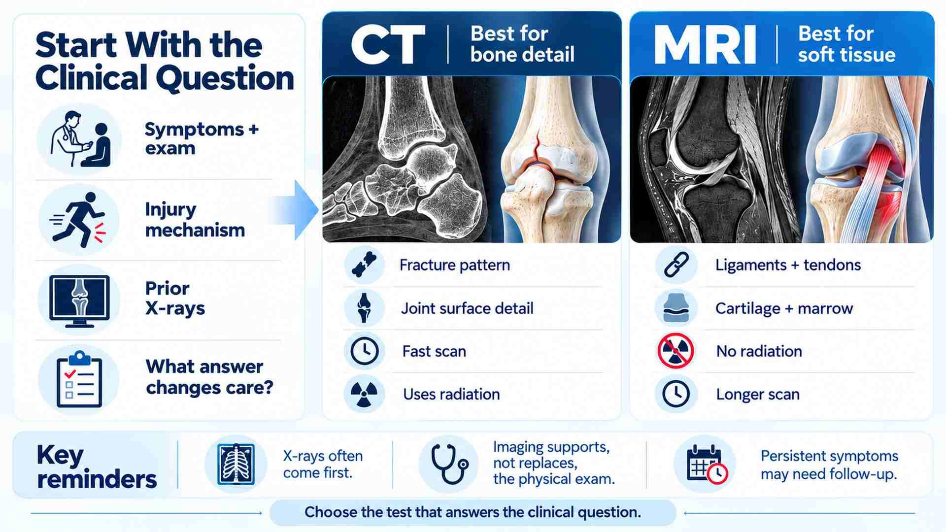
For many suspected fractures and dislocations, X-rays provide the initial information needed to guide treatment. Advanced imaging is considered when the examination or earlier images leave an important question unanswered.
The American College of Radiology’s Acute Shoulder Pain guidance, revised in 2024, recommends radiographs for initial assessment. If a hidden shoulder fracture is still suspected after negative or inconclusive radiographs, either CT or MRI without intravenous contrast may be appropriate. That recommendation concerns a specific clinical situation; it is not a reason to order both scans for any painful shoulder.
CT produces cross-sectional images that can be examined in different planes. It can clarify how bone has broken, whether fragments have moved, and whether a fracture involves a joint surface.
Consider an ankle X-ray that shows a fracture but does not clearly show its relationship to the weight-bearing joint. CT may give the specialist useful detail for planning stabilization or deciding how the limb should be protected. Additional imaging can be useful when it informs a treatment decision.
Routine CT generally provides less detail than MRI about the internal structure of menisci, ligaments, and many tendons. A negative CT may reduce concern about certain fractures without explaining why a knee locks, an ankle gives way, or an arm feels weak.
Those symptoms still need to be considered alongside the examination. A scan that answers one question should not be treated as clearance for an unresolved problem.
MRI may be useful when the concern involves a torn ligament, damaged tendon, cartilage injury, disc abnormality, or a change within bone marrow. It can also identify some fractures and bone injuries that are not visible on initial X-rays. These applications are described in ACR and RSNA’s musculoskeletal MRI guide.
Examples include a knee that locks or gives way after an injury, persistent ankle instability, or difficulty actively lifting an arm after a fall. The clinician uses the history and examination to decide whether MRI would help investigate the suspected structure.
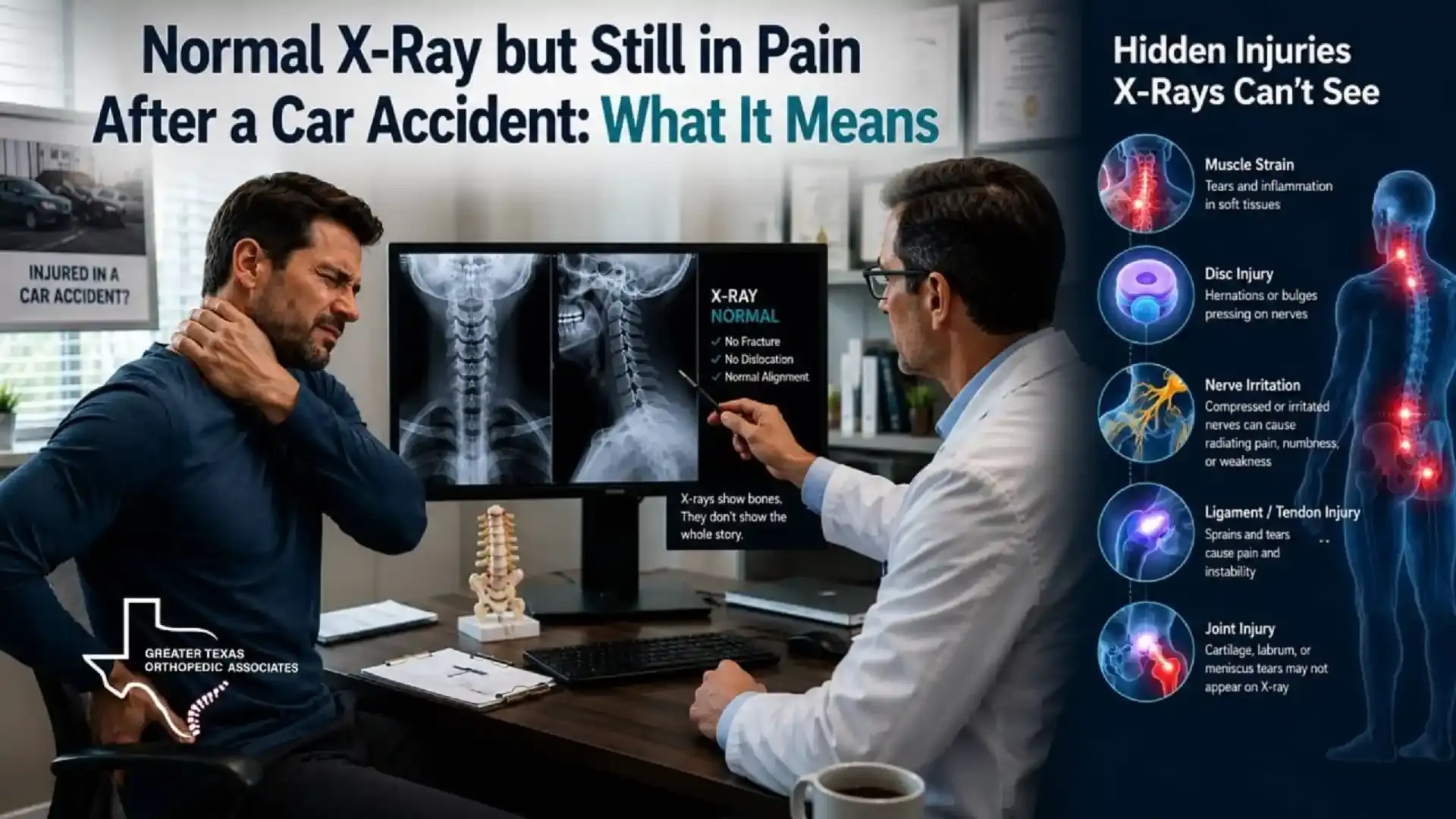
MRI can show bone marrow edema, but edema is a finding that needs interpretation. It does not, by itself, establish the complete diagnosis or the severity of an injury. If initial images do not explain ongoing symptoms, our article on pain despite a normal X-ray after a car accident explains why reassessment may be needed.
MRI is not the only option for a soft tissue question. In selected circumstances, musculoskeletal ultrasound can assess certain tendons and ligaments. Your clinician may recommend a different test if it is better suited to the structure and problem being investigated.
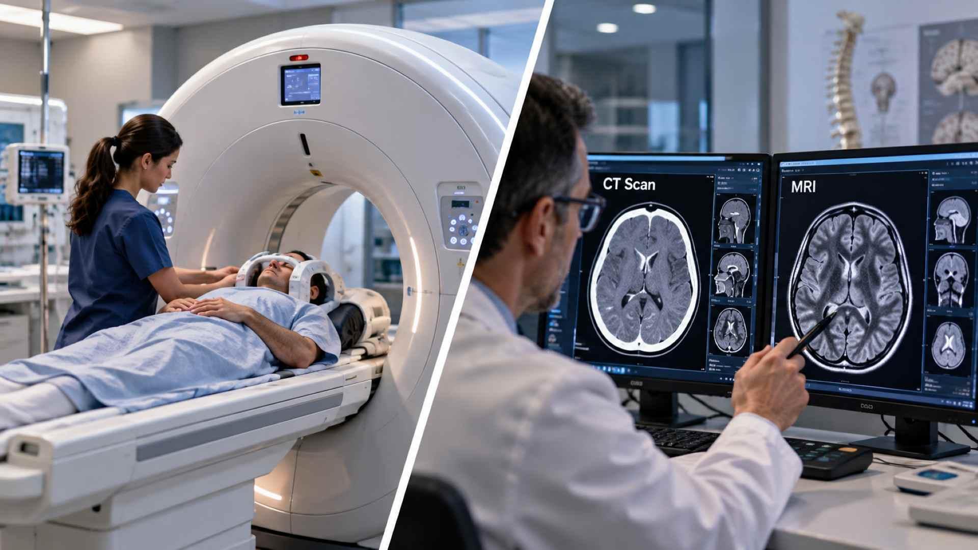
A radiologist reviews your CT or MRI images and prepares a report for the clinician who requested the scan. Your treating clinician then considers the report alongside your symptoms, examination, and previous imaging. Ask who will discuss the findings with you and what they mean for treatment or activity. A report finding alone does not determine the next step in care.
A tear on a report does not automatically mean surgery is needed. Some findings may reflect changes that existed before the injury, while others may be incidental. Your clinician needs to decide whether the finding matches the location of your symptoms and what happens during the examination.
Neither CT nor MRI directly measures your pain or determines how safely you can climb stairs, lift at work, or drive. Imaging also cannot independently establish when every abnormality developed or whether an accident caused it. Symptom onset, previous problems, physical findings, and progress over time all contribute to that assessment.
If the images do not explain persistent symptoms, follow-up may involve another examination, specialist assessment, appropriate protection of the injured area, or a different investigation. Ask what activity is safe while the unanswered question is being assessed.
CT uses ionizing radiation; MRI does not. Tell the imaging team if you are pregnant or might be pregnant so they can assess the examination and any precautions. Concerns about imaging should not delay necessary emergency assessment.
CT examinations are generally quicker. MRI usually requires lying still for longer, and the scanner can feel confined and produces loud sounds. If pain makes it difficult to hold a position, or you experience claustrophobia, discuss this before the appointment so the team can plan appropriate support.
An implant does not automatically prevent MRI. Some devices are MRI conditional, meaning a scan is permitted only under specified conditions. The imaging team must verify the exact device and the conditions that apply. Report implants, metal fragments, and previous procedures even if you have had an MRI before. Metal may also affect image quality.
Many orthopedic CT and MRI examinations do not require intravenous contrast. When contrast is indicated, CT commonly uses an iodine-based agent and MRI a gadolinium-based agent. Tell the team about kidney disease and previous contrast reactions. The suspected problem and examination protocol determine whether contrast would add useful information.
A useful discussion connects the proposed scan to a decision about your care. You can ask:
Bring previous imaging reports and, when available, the images themselves. Explain where the pain occurs, which movements are difficult, and whether symptoms are improving or worsening. Those details help the team select the body region and protocol that address the actual concern.
After significant trauma, a visibly deformed limb, an open wound over a suspected fracture, a cold or pale limb, or rapidly worsening neurological symptoms needs emergency assessment. New bladder or bowel dysfunction with back pain, particularly with numbness around the groin or buttocks or leg weakness, may indicate cauda equina syndrome and requires immediate emergency care. Choosing a scan must not delay that evaluation.
New weakness, increasing difficulty bearing weight, or substantial joint instability warrants prompt clinical review. Persistent milder symptoms that interfere with sleep, work, or daily activity can be discussed at a scheduled visit, with earlier reassessment if they worsen.
The practical question when comparing a CT scan vs MRI after injury is what still needs to be understood. CT may clarify a complicated fracture, while MRI may help investigate a suspected soft tissue or hidden bone injury. Either result needs to be considered alongside your symptoms and examination.
For non-emergency concerns, GTOA’s orthopedic evaluation services in Texas provide context for assessing injuries and discussing an appropriate imaging plan. You can also explore our injury care and specialty services. Emergencies require emergency services rather than waiting for an office appointment.
Neither is better for every injury. CT can clarify complex fractures and joint-surface detail. MRI often provides more information about ligaments, tendons, cartilage, and selected hidden bone injuries. The suspected problem and the treatment decision guide the choice.
No. A scan may answer one question without explaining persistent pain, weakness, or instability. Symptoms that continue or worsen may need reassessment alongside the examination and your functional limitations.
Many orthopedic scans do not require intravenous contrast. Your clinician and imaging team select the protocol according to the suspected problem. Tell them about kidney disease, possible pregnancy, and previous contrast reactions before the examination.
Possibly. The imaging team needs to identify the device and verify whether scanning is permitted under the conditions of the planned MRI. Tell them about implants and metal fragments even if you have previously had an MRI without a problem.
Prepared for Greater Texas Orthopedic Associates. Editorial revision: October 19, 2026. This article provides educational information and does not replace an individual assessment by a qualified clinician.
Comments are reviewed before they appear. For appointments, records, or medical questions, use the contact page.
No comments have been published on this article yet.