Loading article
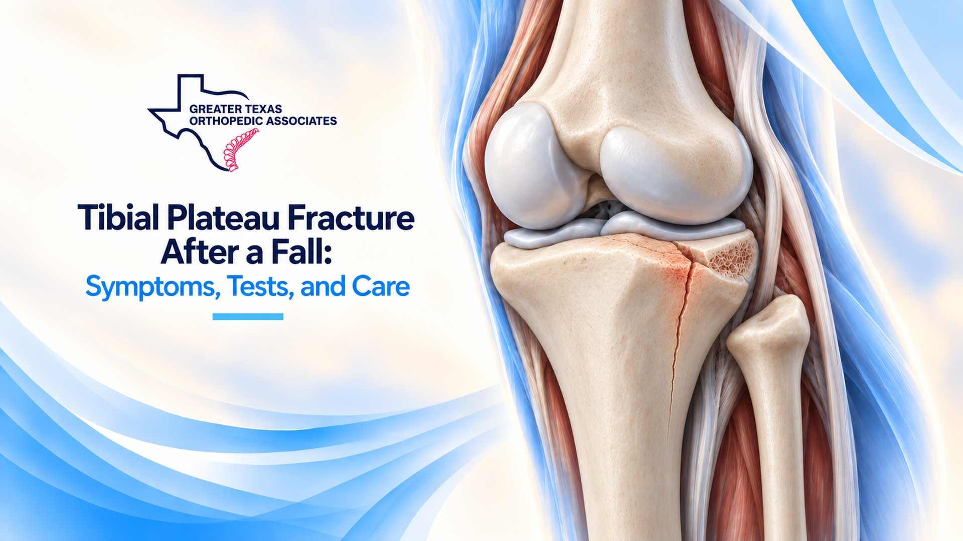
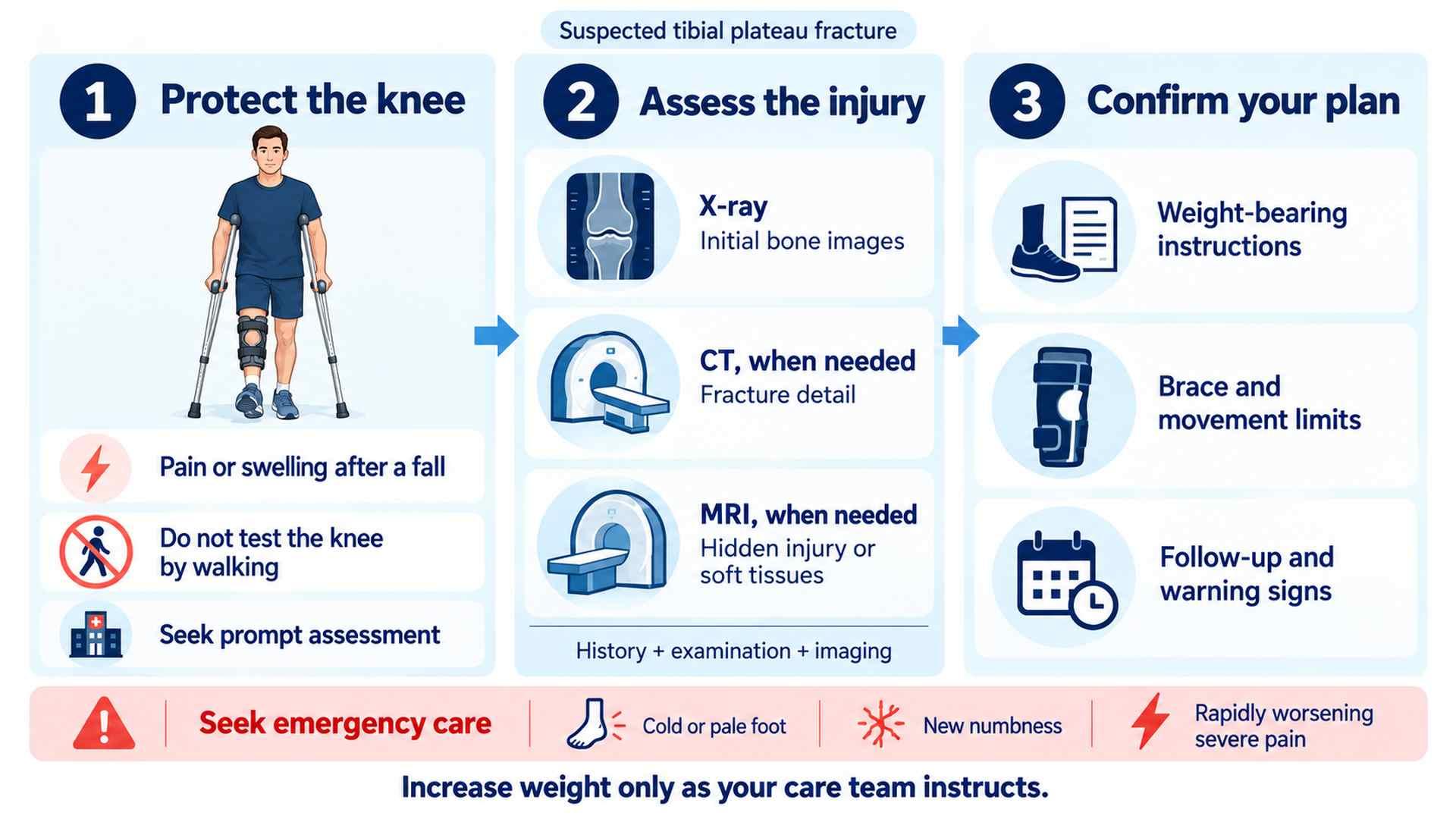
A swollen, painful knee after a fall can leave you wondering whether you have a sprain or a broken bone. A tibial plateau fracture after a fall affects the weightbearing surface at the top of the shinbone. It needs assessment to identify the fracture pattern, check circulation and nerves, and establish safe movement and weightbearing limits. Until you are assessed, avoid repeatedly testing the knee by trying to walk on it.
The tibial plateau is the broad upper surface of the tibia, directly beneath the thighbone. Cartilage covers its joint surfaces, and the menisci help distribute load. A fracture can crack, split, or push part of this surface downward, sometimes affecting alignment or stability.
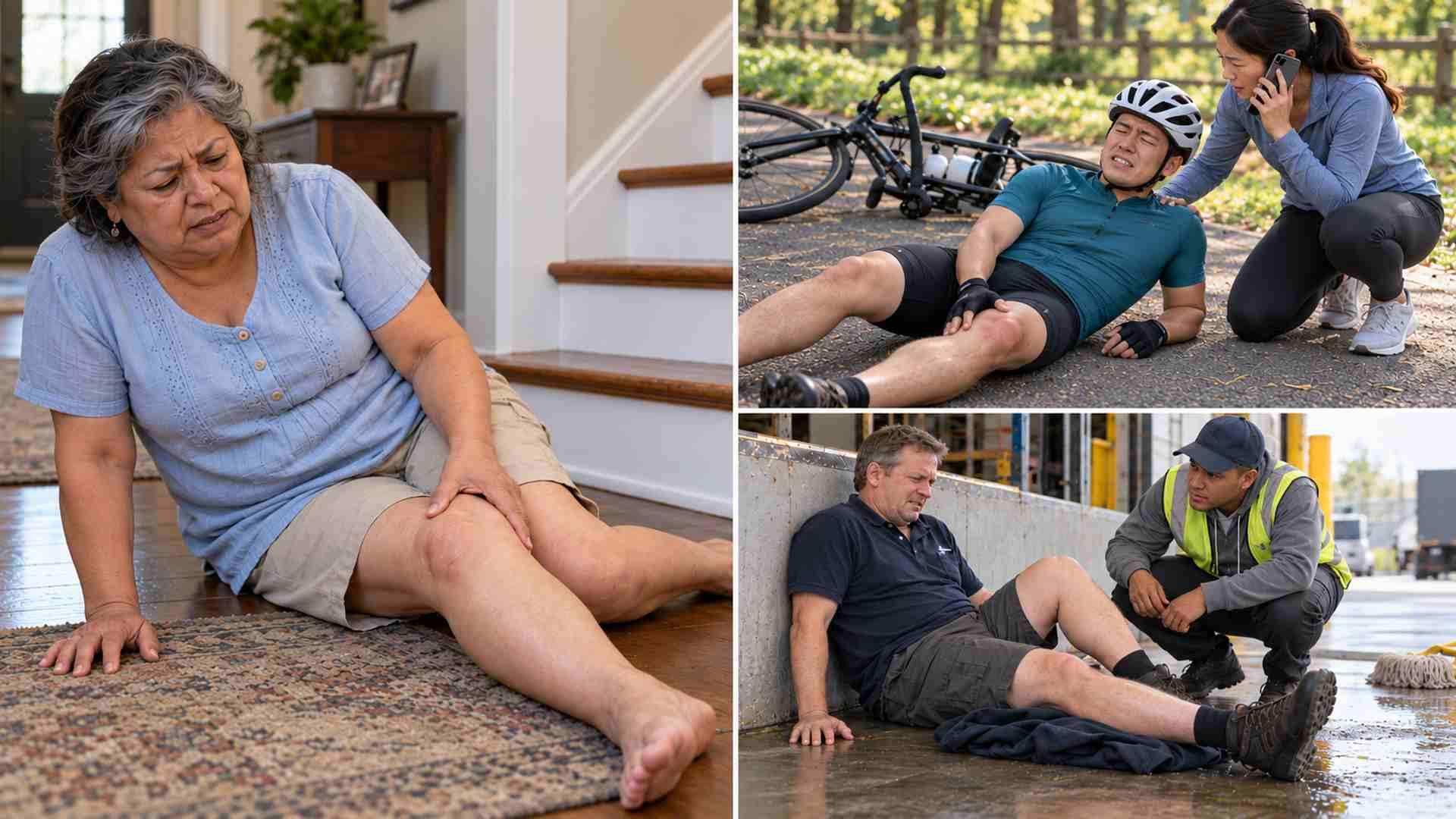
A fall may drive the thighbone into the upper tibia. This can happen after a fall from height or a ground-level slip, particularly when bone strength is reduced. In an older adult injured by a simple fall, ask whether bone health needs assessment. Fall height alone does not establish severity.
The patella, or kneecap, is a different bone at the front of the knee. GTOA’s guide to kneecap fractures after a car accident explains that injury. Pain location helps guide examination but cannot identify the fracture on its own.
Every comment is reviewed before publication. Do not include medical details, personal identifiers, or information about an active claim. This form is not monitored for medical questions or emergencies.
Three more from the same thread.
Showing the first 3 of 3 articles
Typical tibial plateau fracture symptoms include pain near the upper shin or knee joint, swelling, tenderness, limited bending, and difficulty bearing weight. The knee may feel unstable. Some people can take a few steps despite a fracture, so walking ability alone does not establish that the injury is minor.
Tell the clinician how you fell, where the pain is strongest, how quickly swelling developed, and whether you could stand afterward. Mention previous knee problems and report changes in foot sensation promptly.
Circulation problems and compartment syndrome, a dangerous pressure buildup within the leg, can threaten tissue even when the skin is intact. Do not wait for an office appointment when emergency signs develop.
Knee fracture diagnosis combines the injury history, examination, and imaging. The clinician checks tenderness, swelling, skin condition, alignment, sensation, circulation, and how much movement is safe. A sprain, bone bruise, and fracture can cause overlapping symptoms.
Describe whether the foot stayed planted, the knee twisted, or you struck a surface. These details may help the clinician assess the possibility of associated ligament or meniscus damage.
X-rays are usually the starting point when a fall causes focal tenderness, substantial swelling, or difficulty bearing weight. They may show a break or a depressed joint surface. A subtle fracture can be difficult to see initially, so a normal or uncertain X-ray does not end the assessment when clinical concern persists.
CT provides detail about bone fragments and a split or depressed joint surface, which can help with surgical planning. MRI may be useful for a suspected hidden fracture or associated ligament or meniscus injury. These tests answer different questions; you do not automatically need all three. Our guide to CT scan vs MRI after injury explains their different roles.
Nondisplaced means fragments remain in position; depressed means part of the joint surface is pushed down; comminuted means the bone has broken into several pieces. Treatment also depends on stability, alignment, skin condition, and overall health. Persistent severe difficulty walking despite a reassuring report deserves reassessment.
Initial tibial plateau fracture care usually includes restricting weightbearing and supporting the knee with a splint or brace when appropriate. Ask for written instructions about permitted loading, brace use, and knee movement. Nonweightbearing means keeping weight off the injured leg; partial weightbearing needs a specific limit and instruction.
Have someone demonstrate safe use of crutches or a walker. Explain any stairs, bathroom access problems, or difficulty using mobility aids before discharge. An orthopedic evaluation can help clarify restrictions and follow-up needs.
Ask for a demonstration of getting up from a chair and managing the steps you use at home. If you cannot safely reach the bathroom or enter your home, discuss help or equipment before leaving.
A fracture with acceptable alignment, a stable joint, and limited displacement may be treated without surgery. Protection and repeat imaging help check that the position is maintained. Motion may be introduced at a carefully chosen stage to limit stiffness; brace settings and exercises are individualized.
Keep scheduled reviews even if the knee feels better. Repeat imaging helps the treating team check whether the bone fragments have changed position while healing progresses.
Surgery may be discussed when the joint surface is substantially displaced or depressed, the knee is unstable, alignment is unacceptable, or there is important associated damage. The surgeon considers swelling and skin condition as well as the bone. Treatment may involve realigning fragments and securing them with plates or screws. Some complex injuries require staged treatment.
Potential complications include infection, wound problems, stiffness, clots, and fixation failure. Without surgery, displaced fragments or poor alignment may remain a concern. Ask how the benefits and risks apply to your fracture and health. Surgery cannot guarantee a painless knee or prevent future arthritis.
Swelling and soft-tissue injury may affect when surgery can be performed. Ask whether the proposed treatment is temporary protection or definitive fixation, and what follow-up is planned.
Recovery balances protecting the healing joint with limiting stiffness and muscle loss. Supervised motion may begin before weightbearing increases, depending on stability and treatment. Later rehabilitation addresses strength, walking, balance, and everyday tasks. Follow the prescribed exercises rather than a general knee workout.
Recovery is usually measured in months. Some people need three months or longer before full weightbearing is safe; this is not a fixed deadline. Pain relief alone does not mean the bone is ready for more loading. Follow-up X-rays and examinations help the team decide when restrictions can change.
At follow-up, ask what must improve before loading or exercise increases. The team considers fracture position and healing alongside swelling, movement, strength, and your response to exercises. If a visit is delayed, ask whether the current restrictions should continue. Bring specific problems, such as showering or managing stairs, to the discussion.
Driving requires reliable pedal control and quick reactions, without impairment from medication or interference from a brace. Discuss clearance with your clinician. Desk work may resume before work involving ladders or uneven ground. Ask about your actual job, commute, and sport rather than one return date for every activity.
Longer-term concerns can include stiffness, instability, reduced motion, malalignment, and posttraumatic arthritis; these are risks, not inevitable outcomes. Sudden calf pain or swelling warrants prompt assessment for a possible clot. Chest pain or unexplained shortness of breath requires emergency care. Fever, wound drainage, increasing redness, or a sharp decline in function also needs timely medical attention.
Keep your discharge instructions, imaging reports, and treating team’s telephone number together. The GTOA website lists outpatient services and clinic locations for planned follow-up. Emergency signs still require emergency assessment.
Some people can take a few steps despite a fracture. This does not make walking safe. Follow the prescribed weightbearing restrictions.
Yes. Persistent clinical concern may lead to CT or MRI after reassessment, even when the first X-ray appears normal.
No. Alignment, displacement, joint stability, surrounding tissues, and overall health guide the choice between nonsurgical treatment and surgery.
Progress depends on healing, strength, movement, and the activity itself. Ask your clinician for guidance; pain relief alone is not clearance.
A cold or pale foot, new numbness or weakness, an open wound, deformity, or severe escalating pain with tense swelling needs immediate assessment.
For a tibial plateau fracture after a fall, a plan needs to be safe and practical. Explain the instructions back in your own words: how you will move at home, which movements are allowed, and when follow-up is due. Ask for clarification if the restrictions do not fit your circumstances.
Report new or worsening symptoms rather than waiting for a routine review.
This article provides educational information and does not replace an individualized medical evaluation.
For additional patient information, see AAOS OrthoInfo: Fractures of the Proximal Tibia (Shinbone).
Comments are reviewed before they appear. For appointments, records, or medical questions, use the contact page.
No comments have been published on this article yet.